Early Events in Amyloid Formation by Lysozyme Detected by Microfluidic Modulation Spectroscopy
Qiuchen Zheng, Valerie A. Ivancic, Libo Wang, Eugene Ma - RedShiftBio
Noel D. Lazo - Clark University
The self-assembly of lysozyme to form amyloid fibrils is associated with systemic amyloidosis, a disease characterized by the presence of amyloid deposits in various organs of the body. The early events associated in the self-assembly of lysozyme are not well understood. In this work, we used Microfluidic Modulation Spectroscopy (MMS) to characterize the early events in the self-assembly of human lysozyme. Through MMS, we were able to probe the mid-IR absorption band of the protein which is sensitive to both α-helix and β-structure. With heating at 60 ˚C, the β-turn content of the protein increases while its α-helical content decreases. This result suggests that the first structural transition in the self-assembly of human lysozyme is an α-helix to β-structure conformational rearrangement.
Protein Secondary Structure Changes in Amyloid Formation
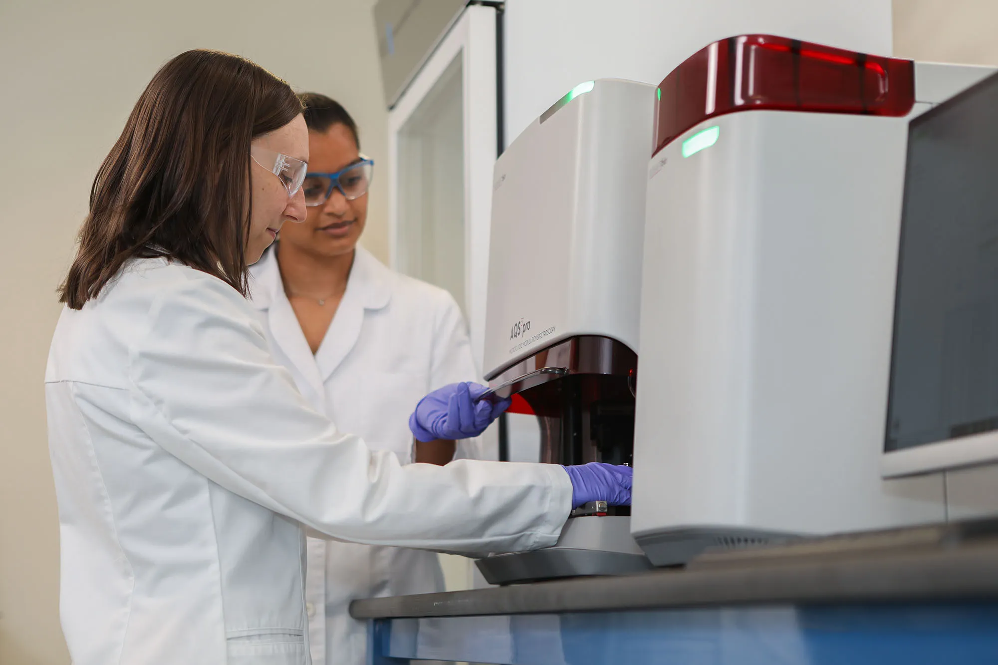
Clark University’s Lazo Lab uses a variety of biophysical techniques to analyze the structure of amyloid proteins. These techniques including circular dichroism, mass spectrometry, electron microscopy and molecular modeling are used to probe the folding and self-assembly of proteins associated with disease. Here, the team used Microfluidic Modulation Spectroscopy AQS3pro (now the Aurora TX) to explore the role of secondary structure changes in the development of amyloidosis.
Analyzing Alpha Helix and Beta Structure
The self-assembly of lysozyme to form amyloid fibrils is associated with systemic amyloidosis, a disease that features amyloid deposits in various organs of the body. The causes of self-assembly of lysozyme are not well understood. In this work, the authors used Microfluidic Modulation Spectroscopy (MMS) to characterize the early events in the self-assembly of human lysozyme. Using the AQS3pro, they were able to probe the mid-IR absorption band of the protein which is sensitive to both α-helix and β-structure. With heating, the β-turn content of the protein increases while its α-helical content decreases. This result suggests that the first structural transition in the self-assembly of human lysozyme is an α-helix to β-structure conformational rearrangement.
Microfluidic Modulation Spectroscopy Compared to Circular Dichroism
Compared to Circular Dichroism (far UV-CD), Microfluidic Modulation Spectroscopy is more sensitive to the early events of amyloid formation by HL and HEWL, primarily because of the sensitivity of the mid-IR region to beta structures.
Clark University’s Lazo Lab also published data using Microfluidic Modulation Spectroscopy AQS3pro looking at the secondary structure of amyloid proteins.


